Difference between revisions of "Pipeleline for simulatneus EEG-fMRI recording"
From BESA® Wiki
| Line 19: | Line 19: | ||
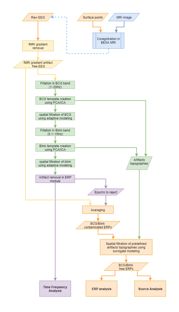
[[File:EEG-fMRI pipeline.png]] | [[File:EEG-fMRI pipeline.png]] | ||
| − | Please note that some steps are grouped with colors: | + | ===Please note that some steps are grouped with colors:=== |
* orange color indicate steps that are part of typical processing of ERP data. | * orange color indicate steps that are part of typical processing of ERP data. | ||
* blue color indicate optional, yet strongly recommended EEG-MRI data co-registration. | * blue color indicate optional, yet strongly recommended EEG-MRI data co-registration. | ||
| Line 25: | Line 25: | ||
* green steps are reserved for BCG/blink artifact correction | * green steps are reserved for BCG/blink artifact correction | ||
* violet color indicate steps for time frequency analysis. Here also information about rejected epochs is provided for averaging. | * violet color indicate steps for time frequency analysis. Here also information about rejected epochs is provided for averaging. | ||
| + | |||
| + | ==fMRI gradient removal== | ||
| + | To remove fMRI gradient go to | ||
Revision as of 18:06, 9 April 2018
| Module information | |
| Modules | BESA Research Basic or higher |
| Version | 7.0 or higher |
Contents
Pipeline for simultaneous EEG-fMRI recording
Before you start
- You have jitter between trials in ERP experiment (i.e. random value of ±200ms). Some further guidelines about paradigm creation can be found here: (Rusiniak et al., 2013a).
- The subject movement is limited to minimum.
- Electrode to skin impedance is as low as possible.
- EEG-fMRI recording session was long enough to allow for proper artifact creation. Usually experiment should last at least 6 minutes.
- Especially for first few registrations repeat the experiment outside of the bore to compare results.
Pipeline overview
The recommended pipeline of processing EEG data registered during fMRI session looks as follows:
Please note that some steps are grouped with colors:
- orange color indicate steps that are part of typical processing of ERP data.
- blue color indicate optional, yet strongly recommended EEG-MRI data co-registration.
- yellow color marks the steps related to fMRI gradient artifact removal
- green steps are reserved for BCG/blink artifact correction
- violet color indicate steps for time frequency analysis. Here also information about rejected epochs is provided for averaging.
fMRI gradient removal
To remove fMRI gradient go to
